An essential piece of equipment
Without a microscope it is simply impossible to tell the difference between a water quality and a parasite problem. The microscope should be considered the most basic of tools in fish disease diagnosis – indeed, an accurate, full diagnosis of the disease and its cause just isn’t possible without a microscope!!

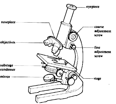
A modern monocular microscope
Why is it so important?
Unusual behaviour such as heavy breathing, rubbing, flashing, lethargy is often taken as a sign of parasite disease – yet the same behaviour can be due to water quality problems, internal organ disease or many other causes. Without a microscopic examination of both the skin and gills it is simply impossible to tell the difference and any treatment undertaken is based on nothing more than guesswork! While simply taking a guess as to the cause and treatment required may work it is just as likely to make matters worse. So microscopy is a vital and basic step in diagnosing and treating fish disease.
Buying a microscope
They come in a wide range of types and prices, costing less than a hundred to several thousand pounds – although fish keepers generally do not require an expensive model. There is also a market for good second-hand ‘scopes.
It really depends on what you want to use it for. Most of the common parasites such as flukes, white-spot and Trichodina can be easily identified with the cheaper models, but a better quality model is required for critical examinations of cell structures and some small parasites. Generally speaking as prices increase you are paying for better engineering, illumination and optics
Models for fish keepers
There are two basic styles of microscope available. The cheaper monocular model has a single viewing tube (as above) – which is fine for occasional use. The binocular models enable you to view with both eyes, giving a better field of view. If you want to take photos or video – then you will really need a trinocular model with a dedicated phototube.
Two other considerations which can make a considerable difference are illumination and the stage.
Illumination
The better the lighting – the clearer the image. Most of the cheap models have an understage mirror which reflects light to illuminate the slide. This can pose problems in dull conditions or lead to contortions with table lamps to try and improve the illumination of the specimen being studied. By far the best option is a fixed or plug-in understage light system, which gives a consistent amount of light. A basic plug-in system can be purchased for under £50 and, in my opinion, is money well spent. More expensive microscopes have built-in halogen lamps with brightness control
Stage
The stage is the part of the microscope where the slides are placed for viewing. As you will appreciate, only a small part of the slide can be seen at any time and the slide needs to be moved to see other parts. Incidentally, the view that can be seen at any time is called the field of view, which reduces at higher magnifications.
On the cheaper models the slide is held by clips and has to be moved manually – but this cannot be done smoothly and it is virtually impossible to return to a given position on the slide.
All but the most basic of models have a mechanical stage fitted with vernier screws that allows you to move the slide smoothly and examine it in a methodical pattern. Again, this usually adds less than £50 to the basic cost and is invaluable when scanning the slide for parasites.
There are different qualities of mechanical stage- and some of the more robust stages can be quite expensive.
