Click the right-hand mouse button if you want to down-load any of the movies (click “save target as….). The file sizes have been kept reasonably small, hopefully without losing too much detail – they will take a minute or two to download- depending on your modem speed. Let them fully download before playing them. If you are using Windows Media Player, click the pause button and then you will see when the movie has fully loaded. All videos are in mpeg format.
 Argulus 01 (fish louse) (285 kb) High power magnification giving a fish’s view of this nasty parasite. It shows the highly flexible suckers that the louse uses to attach to the fish’s body. In between the suckers you can see a large, flexible proboscis-like mouth that the louse uses to suck out the fish’s body fluids – nice!
 Argulus 02 (fish louse) (213 kb) Low power view of Argulus (x100) – showing egg-sac, legs, suckers etc.
 Chilodonella 01 (315 kb) Shows Chilodonella at 100 x magnification. The parasite is flat, oval to heart shape with a notched anterior end. Characteristic gliding action, moving slowly and turning in circles. Flattened shape can be seen when the parasite turns sideways.
 Chilodonella 02 (430 kb) Shows Chilodonella x 400 magnification in phase contrast. You can just about see the characteristic bands of cilia
 Ichthyobodo – (Costia 01) (373 kb) Costia shown at 600 x magnification. A very fast moving parasite. Characteristic flickering motion as it turns its crescent-shaped body.
 Dactylogyrus 01 (Gill fluke) (212 kb) A gill fluke on the edge of a gill filament at 100x magnification. Characteristic black eye-spots with scalloped head. You can see trichodinids in the background.
 Dactylogyrus 02 (Gill fluke) (414kb) Magnification x600 showing fluke being parasitised by trichodinids – obviously dog eat dog!
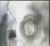
 Dermocystidium 01 (306 kb) An uncommon fungal-type disease that affects koi. Note the intact immature nodules and the ruptured lesions showing white hyphae.
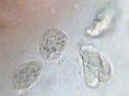
 Gyrodactylus 01 (skin fluke) (378 kb) Gyrodactylus can be differentiated from Dactylogyrus by the absence of eye-spots. This movie shows Gyrodactylus at 100x magnification. Note the embryo clearly visible in abdomen (the hooks of the embryo are visible). Also note the arrangement of attachment hooks at rear end (opisthohaptor).
 Gyrodactylus 02 (skin fluke) (325 kb) Gyrodactylus at x 600 magnification shows the fearsome arrangement of hooks on the opisthohaptor together with a pair of hooks in the abdomen – the yet-to-be-born embryo! No wonder fish flash and jump!
 Trichodina 01 (from the gill of a koi) (358 kb) High power (x600) shows trichodinids grazing on the gill filaments of a koi. Note the whirling cilia (minute ‘hairs’ used for motion and to fan food into the mouth). Typical ‘flying saucer’ motion. Gill trichodinids are usually smaller than skin-dwelling trichodinids (<30 µm).
 Trichodina 02 (from the skin of a koi) (112 kb) Not a lot of movement but it shows a typical wet mount of a mucus scrape x 100.
 Ichthyophririus multifilis (white spot 01) (376 kb) Shows typical wet mount at x100 magnification. Large dark parasites (trophonts) with typical horseshoe macronucleus clearly visible. White spot has a lazy rolling action. Note the variance in size of the trophonts.
 White spot 02 (334 kb) Higher magnification and phase contrast (x600) shows rows of cilia beating, providing means of motion. Whitespot is a holotrich ciliate – the cilia are distributed evenly over the entire body surface.
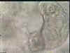
 Plumatella 01 (283 kb) Plumatella sp. A freshwater Bryozoan (moss animal). A shy, charming little animal that lives in some filters. It is a beneficial organism that lives on smaller protozoa, bacteria, algae and just about anything it can trap with its tentacles.