Practice make perfect
Although the microscope is a fairly simple instrument to set up and use, interpreting what you see can sometimes be difficult. It really requires practice until such times as you know what is normal and what is not.
As a piece of advice I would suggest familiarizing yourself with healthy or non-urgent fish mucus samples – rather than waiting until a serious case arises. By getting used to taking and examining mucus samples you will be better prepared to make a judgment when a serious problem arises.
What to look for
I am assuming that initially the microscope is going to be used as as simple diagnostic aid to carry out procedures such as skin and gill examinations. It can of course be used for more advanced studies, such as plant and animal cell structure. You will probably have seen photos of slides on the site showing details of various fish organs such as gills. This is histology and involves special preparation and staining of the specimen.
At a basic level, microscopy would be used as part of a routine examination to check mainly for external parasites. This involves taking a skin or gill scrape / sample and making a simple wet mount as previously described.
Initially it is very easy to get confused by what you see on the slide – particularly if small non-parasitic aquatic animals happen to be sampled, leading to fears of some new, frightening parasite or disease!
Normal mucus
The first stage is to recognize what normal mucus looks like and disregard any “debris” or unimportant “introductions”. These could be things like air bubbles trapped under the cover slide which appear as circular, dark-rimmed “grommets”. Or perhaps normal cellular debris which appears as stationary, irregular-shaped – often dark patches. You are also likely to see trapped algae of all shapes and sizes. This is why it is important to get as much practice as possible before using your microscope in earnest.
|
Normal mucus in a wet mount at 100x magnification |
|
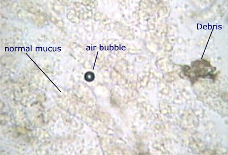 Normal mucus which is seen a lumpy, transparent, light to grey substance. Also shown is a trapped air-bubble which is often mistaken for a strange parasite, and some typical cellular debris. Active parasites should be easily seen. Normal mucus which is seen a lumpy, transparent, light to grey substance. Also shown is a trapped air-bubble which is often mistaken for a strange parasite, and some typical cellular debris. Active parasites should be easily seen. |
|
|
photo: Frank Prince-Iles |
|
Five parasites
At the risk of oversimplifying what can be a complex issue, in the vast majority of cases (>90%), the usual findings would be a common parasite problem. In freshwater fish such as koi and goldfish these usually involve just five different types namely, flukes, Costia, Trichodina, Chilodonella or white-spot. So, simply being able to recognize these common beasties will make you a reasonably competent microscopist. There are, as already suggested, less common things you might come across, but once you are familiar with these basics, it is fairly easy to focus on any abnormal findings and either look them up in a good book or ask for advice.
In most slide preparations the parasites will be “alive and kicking” and so they are easy to spot and recognize. However, it is important to scan the entire slide, slowly and methodically looking for parasites that are still. These can be more difficult to spot as all of these parasites are transparent and therefore tend to blend in with the mucus. However, with the exception of Costia, which is virtually impossible to spot unless it is moving, they can be seen once you have the experience and have “got your eye in”.
This is why it is important to examine slides as soon as they are prepared as many parasites die if left for any period.
Recognizing parasites
There are many good books available which describe the common parasites. There are further details and photos on the relevant pages of this site. The movies of parasites will also give some idea of the way each parasite moves; an important diagnostic feature. I have included a short summary of each of the parasites below, but you will need to check the relevant pages for more detail;
- White-spot: Dark, slowly rotating circular parasites of varying sizes. Often with a lighter, horseshoe-shape visible. One of the larger parasites. Go to page
- Flukes: Long, worm-shaped animals that move in looping action. There are often clearly visible hooks at one end. Go to page
- Trichodina: Medium=sized round parasites with a series of inner, concentric rings. These zoom around like flying saucers. Go to page
- Costia: The smallest parasite that is often easy to miss. A fast-moving parasite recognized by its flashing and twinkling as it moves in and out of focus Go to page
- Chilodonella: A medium-sized oval-shaped parasite that turns and glides. Go to page
Go to fish disease movie clips
Happy hunting!